

DIAGNOSIS
FAHR’S DISEASE
FINDINGS
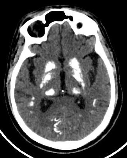
Near symmetric patchy calcifications are seen in bilateral cerebral and cerebellar hemispheres including:
•Bilateral basal ganglia
•Thalami
•Dentate nuclei
•Bilateral corona radiata.
DISCUSSION
Fahr’s disease, also known as bilateral striatopallidodentatecalcinosis, is characterized by abnormal vascular calcium deposition, particularly in the basal ganglia, cerebellar dentate nucleiand white matterwith subsequent atrophy. It typically presentsin 40 to 60 years of age
•Primary: equivalent to familial cerebral ferrocalcinosisor primary familial brain calcification (now the preferred term), with no underlying other cause. Its usually autosomal dominant.
•Secondary: due to an underlying metabolic, infective or other cause
The condition that has been closely described with diffuse, bilateral, symmetric striopallidodentatecalcinosis is primary hypoparathyroidism,lupus, tuberous sclerosis, Alzheimer’s disease, myotonic muscular dystrophy and mitochondrial encephalopathies. When there is no explainable cause for striopallidodentatecalcinosis, the condition is termed as Fahr’s disease.
The treatment of Fahr’s diseaseis directed to the identifiable cause especially hypoparathyroidism. In other cases, symptomatic or conservative therapy with clinical follow-up is the rule.Prognosis is variable, cannot be predicted and is unrelated to the extent of calcification. Death usually occurs secondary to neurological deterioration.


