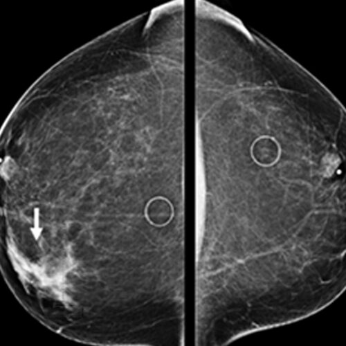
We are happy to announce our expansion of tele reporting in the field of PET CT and Conventional XRAY Mammography with the help of experts in the field of oncoimaging.
There is mild hypertrophy of the basal and mid-myocardial inter-ventricular septum (IVS). It measures 11 mm in mid segment (diastole) and 14 mm in systole.
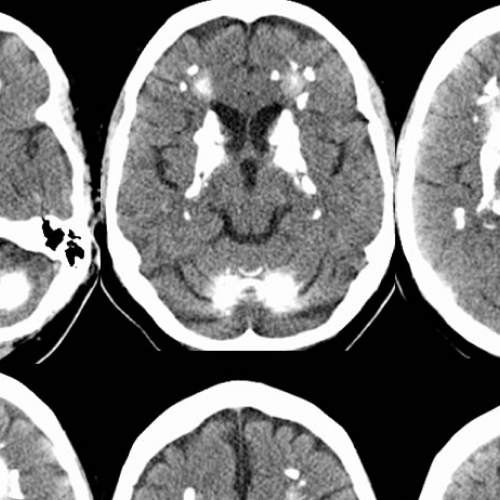
Fahr’s disease, also known as bilateral striatopallidodentatecalcinosis, is characterized by abnormal vascular calcium deposition, particularly in the basal ganglia, cerebellar dentate nucleiand white matterwith subsequent atrophy.
Multiple tiny T1 and T2 isointense nodular lesions are seen along ependymal lining of both lateral ventricles predominantly in the body region probably representing sub ependymal hemartomas.
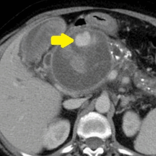
A well-defined oval cyst isseen abutting the pancreatic body and tail, suggestive of a pancreatic pseudocyst. The pancreatic duct is minimally prominent. Thecyst is seen anteriorly abutting the stomach wall with resultant diffuse oedematousthickening of the walls of the stomach.A few ill-defined hyperdensitiesare seen within the cyst, suggestive of chronic hemorrhages/ sludge
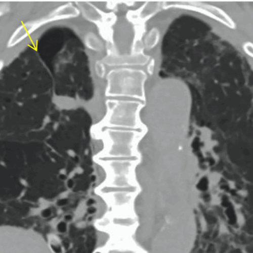
An azygos lobe is a rare normal anatomic variant of the right upper lobe, first described by Heinrich Wrisbergin 1778 due to invaginationof the azygosvein and pleura during development in the fetus.
NON HODGKIN>>> HODGKIN •ATYPICAL, MORE AGGRESSIVE, POOR PROGNOSIS •EXTRANODAL DISEASE MORE COMMON THAN SEEN IN NORMAL INDIVIDUALS •NOT RELATED TO CD4 COUNT BUT SEEN IN MID /LATE PHASE
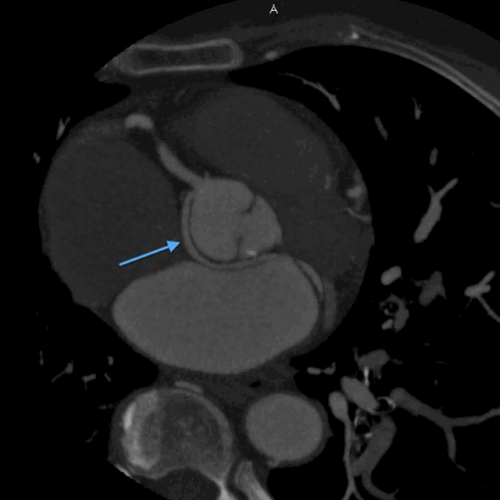
On MDCT angiography, left coronary artery was seen to arise from the left sinus of valsalva which further coursed along the anterior interventricular groove as LAD. RCA was seen to be arising from anterior aortic sinus with its normal course along right atrio-ventricular (AV) groove.